
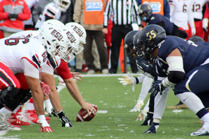
Feb 5, 2024New study suggests brain changes in football players who are subject to head impacts
A study published recently in the JAMA Network Open involving MRI research that adolescent football players are susceptible to a variety of brain changes due to head impacts from playing.
Researchers reviewed findings from structural MRI for 205 high-school football players and 70 high-school athletes who participated in non-contact sports such as swimming and tennis. The study authors also assessed resting-state functional MRI scans for 149 football players and 59 participants from non-contact sports.
 In comparison to athletes in non-contact sports, the study authors noted the MRI findings for football players revealed deeper sulcal depth in “widespread brain regions,” including the frontotemporal regions, precentral gyrus, and the cingulate cortex.
A recent article from Diagnostic Imaging detailed the study on brain changes in football players.
Below is an excerpt from the Diagnostic Imaging article.
“These findings shed light on the potential impact of mechanical stressors on brain structure and provide valuable insights into the dynamics of cortical changes in individuals engaged in football,” wrote lead study author Taylor Zuidema, a Ph.D. candidate who is affiliated with the Department of Kinesiology and the Program in Neuroscience at Indiana University in Bloomington, Ind., and colleagues.
Football players also had cortical thickening in the anterior and posterior cingulate cortex as well as cortical thinning in multiple brain regions, according to the researchers.
“ … Our data showed significant cortical thinning in the frontal-occipital regions of adolescent football players’ brains, accompanied by cortical thickening in the cingulate cortex. TBI and repetitive exposure to head impacts can emerge as potential accelerators of age-related cortical thinning in these identified regions,” explained Zuidema and colleagues.
Three Key Takeaways
1. Structural changes in the brains of adolescent football players. The MRI findings indicate that adolescent football players exhibit deeper sulcal depth in various brain regions, including the frontotemporal regions, precentral gyrus, and the cingulate cortex. This suggests that exposure to head impacts in football may lead to structural alterations in the brain, potentially accelerating tissue atrophy.
2. Cortical thinning and thickening patterns. The study reveals cortical thinning in the frontal-occipital regions and cortical thickening in the anterior and posterior cingulate cortex of adolescent football players’ brains. These patterns may be linked to traumatic brain injury and repetitive head impacts, suggesting potential accelerators of age-related cortical thinning in specific brain regions.
» ALSO SEE: Study reveals longer-term impact on sports-related brain injuries