Oct 11, 2019A comprehensive approach to concussions
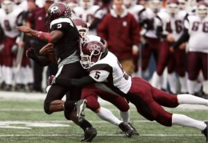
Many athletes assume that a blow to the head is just part of the game. As they may have done many times before, they “shake it off” and resume play. Nobody wants to let down the team.
The last decade has seen a 60% rise in sports-related head injuries (SRC) in emergency rooms (ED) across the country. The steep rise may be attributed to better awareness of deficits, but also to improved accessibility to technology.
The first indication of a possible concussion, and, therefore, the need for intervention and management, is the suspicion of injury, symptoms and symptom provocation. There is no MRI, CT scan or blood test, that as of yet, can diagnose a concussion. In many cases, the hit can be something that initially seems ordinary or unremarkable. The intent is not to discourage play but rather to enable student athletes (0-100 years of age) to engage in sport and recreational activity with better knowledge to keep them playing longer, smarter and safer.
 It’s from this initial injury that student-athletes display symptoms that can take minutes, hours or even days to emerge. Poor sleep or loud noises can stimulate those symptoms. Suspicion of injury, followed by symptoms and symptom provocation, has been the avenue for diagnosing a concussion.
Symptoms can go largely unnoticed, and the significance may be hidden for hours or longer. It’s for this reason that, in our practice, we make our office available to examine these individuals immediately to capture initial signs and indicators.
Establishing baselines
Establishing the baseline of function is critical in developing a complete assessment, points of improvement and future treatments.
The five critical baselines that we address are:
- Vision. Determining acuity, blind spots, blurring, double vision, oculomotor — fixation, pursuits and saccades, near point of convergence (NPC) — using eye tracking software like RightEye.
- Vestibular. Any reports of dizziness, stumbling, falls, nystagmus (eye movements) or vertigo. Also, vestibular-ocular reflex (VOR) assessment and vision motion sensitivity (VMS) assessment using RightEye.
- Balance. Addressing changes in gait or the ability to perform activities — climbing stairs, stepping over objects or walking on uneven surfaces. Conduct a BESS test with eyes open/eyes closed, shoulder-width and in tandem stance.
- Cognition. Including memory, orientation to current events, and problem solving utilizing web-based products such as ImPACT testing. Also, CNS vital signs and CBS health assessment.
- Reaction time. Can be done with or without technology, utilizing Senaptec or DynaVision D2 technology.
We designed our assessment and treatment approach with a multidisciplinary best practice approach. In assessing the effects of concussions and potential consequences, both long and short term, we utilize a team approach. Our professionals include a physical therapist, occupational, athletic trainer and a neuro-optometrist. We find this to be extremely beneficial in testing and interpreting results. As a group, we’re able to create a picture of how the person is functioning and any conditions or effects that may be of concern.
In the beginning evaluation, we establish the system conditions that present at that point in time, a snapshot of the conditions in that moment. By documenting the neurological status, we determine an important baseline. With this information, we can then monitor changes that may occur in the future.
Examining symptoms
Many individuals assume that the initial common symptoms of nausea and headache are the only concerning factors, or proof, of a concussion. It’s important, therefore, to make our initial evaluation and ongoing monitoring a more in-depth explanation of the interwoven brain function that may seem slight and unrelated.
The person may report blurred vision, double vision or sensitivity to light. They may explain feelings of dizziness, and vision similar to stepping off a roller coaster.
Balance problems may be minor, yet connected to a concussion and brain injury. They may notice that they cannot stand on one leg and kick a ball as easily, or that they fall more in gymnastics or skateboarding. Advanced motor skills such as climbing stairs or hiking may seem more difficult. These are frequently missed as symptoms of a previous concussion.
Students can complain of difficulties remembering school lectures and assignments. Individuals may notice moments of confusion that are unusual, like not knowing when or where their next class is scheduled. Fatigue and difficulty sleeping, or sleeping too much, also are subtle symptoms that are often contributed to other causes.
Post-traumatic stress can be another puzzling and underreported pathology. The athlete may appear easily frightened, anxious and withdrawn.
Depression and anxiety also could be attributed to the injury, particularly if they are new to the person. Migraines, as well as neck and back pain, can appear and may be indicative of serious, possibly advancing conditions. Linking them to the incident can help in the diagnosis and treatment of these cases.
Why the eyes
- 70% of our brain is dedicated to vision in some fashion.
- 80% of all sensory goes through the eyes.
- 90% of individuals that have a concussion will demonstrate one or more ocular difficulties. If not addressed, it can result in delayed recovery.
- 40% of individuals will have ocular difficulties longer than three months.
Intervention is helpful in ensuring resolution of ocular complaints and meeting the other pathways. Here are some impairments we look out for:
→ Blurred or fluctuating vision. Deficits in eye focusing (accommodative dysfunction), or a reduced ability to compensate for a minor prescription can result in blurred or fluctuating vision.
→ Double vision. More than half of concussion patients see double. Oftentimes, double vision is only present intermittently, and patients may not realize that what they are experiencing is double vision. One major cause of double vision is convergence insufficiency, or a reduced ability to use the eyes together during near activities.
→ Eye tracking deficits. Difficulties with reading or computer work is extremely common after a concussion. This is one reason why we use eye tracking tests to help diagnose concussions.
→ Light sensitivity. Many concussion patients find that they are bothered by certain types of light, even when indoors.
→ Reduced cognitive abilities. Feeling as if they’re “in a fog” may be a common occurrence. There also can be difficulties with concentration, memory and thinking quickly.
→ Balance difficulties/dizziness. The visual system has a strong influence on balance. When vision conditions are present, it can affect balance and create dizziness and nausea.
→ Headaches. Evaluation of the eyes should be considered when headaches are present, especially when they occur at the end of the day or after visual activities, like reading or computer work.
Vision is multimodal activity of the brain and body
The focal/central vision mode occurs primarily as a macular function. Detail discrimination relates to visual acuity. Other functions within this area include attention, concentration, and orientation to present consciousness. Slow speed of processing is within the occipital cortex. Higher processing is primarily under cortical interpretation/action.
 The periphery/dorsal vision functions can be seen individually and in complex combinations. This is not only important in initial baseline determination, but also in the ongoing evaluation and monitoring. Spatial orientation is required for posture and balance, and the feedback loop of maintaining aligned posture and balance.
Movement depends on visual perception and orientation of space in relation to visual planes. Movement requires a shifting or planning of the constant requirements of posture, balance and orientation.
» ALSO SEE: Examining subconcussive hits in sports
Anticipate change in the preconscious and rapid speed in processing. This can be framed within the “fight or flight” survival actions, as they require fast analysis and response in an almost automatic manner.
We know that 20% of the eye’s nerve fibers do not go to the occipital cortex, but to the midbrain, which delivers this sensorimotor function. Spatial visual processes include preconscious and proactive actions, and reception of cortical feedback.
The complexity of neuro-organization possibilities is structured to move the person beyond a static position or posture, smoothly responding to constant change. When this loop and progression fails, the patient can become immobile and isolated.
What this means is that the balance and interaction between vision and motor is compromised. Vision dysfunction causes recovery delays, interferes with learning, creates problems in communication, and effects memory by disrupting time and space. This causes “focal binding.”
Focal binding compromises preconscious/proactive relationships between the dorsal system (peripheral/motor), vestibular and proprioception. Movement becomes conscious (top down), and reduces function and fluency. There is no fluency, because the systems are no longer able to anticipate (e.g. reading, walking in a crowded space).
Essentially, focal binding inhibits the release of “detail,” with the environment becoming overstimulated. In essence, these individuals can’t see the forest for the trees. Movement in the environment becomes chaos to the visual system. Print on a page becomes a mass of detail, and movement of the eyes is projected into the field, causing movement of print or the perception of movement of the ground being walked on. Focal-binding is why you never start with pencil push-ups — it makes the student athlete worse.
Rehab: Vision and vestibular together?
Cognitive issues that linger show up as unresolved visual vestibular issues. Anxiety problems show up as a consequence of unresolved vision vestibular issues. Cervical issues are more prevalent than previously reported, and they linger because of unresolved vision vestibular problems.
We found that eye tracking is an excellent screening tool. It enables us to help identify potential problems in the vestibular and ocular motor conditions:
- RightEye: High tech.
- VOMS, KD, NSUCO: Low tech eye tracking.
Post-injury assessment
Here is a breakdown of our post-injury assessment:
- History and clinical interview.
- Self-reporting symptom assessment. We use the post-concussion symptom survey(PCSS).
- Vision. Acuity, with a refraction, looking for blowout fractures, iritis, due to trauma. Looking for detached retina.
- Vision/vestibular/balance. We are using VOR testing and VMS sensitivity. RightEye with eye tracking software.
- Cognitive assessment. ImPACT testing, CNS Vital signs or CBS Heath.
- Heart rate variability (HRV). To understand HRV, we first need to understand our nervous system and heart rate. We can trace heart rate variability back to our autonomic nervous system. The autonomic nervous system regulates important systems in our body, including heart and respiration rate and digestion. The autonomic nervous system has a parasympathetic (rest) and a sympathetic (activation) branch. HRV is an indicator that both branches are functioning — the parasympathetic, in particular.
- Reaction time. This is the length of time it takes to respond to a stimulus. Reaction time is important when driving, playing sports, during emergencies, and in many day-to-day activities. Reaction time depends on nerve connections and signal pathways.
Treating the athlete
Once we’ve completed our testing and established the athlete’s condition, this is how we proceed with treatment during the first three days.
- Rest. Rest is still the cornerstone of recovery — we must get them sleeping. We have them take magnesium 30 minutes before they go to bed.
- Diet. Reduce gluten and casein in their diet. These two categories of food are the most irritating to the system. Studies show that within five to 15 minutes of a concussion the blood-brain barrier opens up, as well as the gut barrier. If these barriers do not close up in a timely fashion, this leads to a prolonged recovery. We need athletes to get more protein, reduce the intake of sugar and caffeine, and stay clear of alcohol.
- Planning & pacing. Depending on symptoms and presentation, we must help the athlete understand that “pushing through” can make them worse. They must plan and pace their day. We offer suggestions for school/work attendance, along with planning and pacing.
- Reduce internet/phone/computer time. No more than 10 to 15 minutes of screen time during each hour.
- Therapy. After the assessment, they do 10 to 20 minutes of light therapy; either photobiomodulation or syntonics.
- Epley maneuver. They perform this maneuver, as long as we clear the neck.
- Cranio-sacral therapies. Do this for the head and neck.
- Cervical evaluation. Icing and taping as needed.
Matrix therapy
We will see the athlete every day until the symptoms are down — sleeping better, ROM on the neck, RightEye or VOMS better, HRV shows parasympathetic and sympathetic system are more balanced.
Then, we start the exertion protocol to get them moving. We conducted a study of 200 concussions that presented at zero to seven days of the injury. In the self-reported symptom survey, we averaged 19% to 24% reduction in symptoms with 45 minutes of therapy — light, cranio-sacral and Epley maneuver. We are working to publish it.
It takes a team to help student-athletes of all ages recover from a concussion. Vision, vestibular and cervical are the pathways to start with, along with understanding the parasympathetic-sympathetic system with HRV. We must get athletes sleeping, and make sure they’re getting good protein, and reducing sugars and caffeine in their diet. If there is success, anxiety, post-trauma migraine, and cognitive issues can be better resolved.


